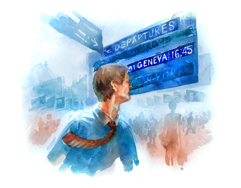
I’m late! Excuse me, is it a plane to Geneva or to Gdansk?
See what you want, when you want. Laser surgery will free you from unnecessary stress.
For those who do not want or can not wear glasses, medicine has found the perfect solution – refractive surgery. It is a new direction in eye microsurgery designed to reduce or completely remove vision defects. The most effective and safe way of correcting treatments using an excimer laser.

Methods
FemtoLASIK
is a method of laser vision correction using the most technologically advanced femtosecond laser. This laser is used in the initial phase of LASIK surgery to create the corneal flap. This type of laser is more accurate and predictable than the conventional procedure using the microkeratome. Using femtosecond laser surgeon can adjust diameter, thickness and position of flap. The computer program arranged to create a flap for each patient individually. The incision is programmed and carried out to an accuracy of thousandths of a millimeter.In FemtoLASIK greatest advantage over traditional LASIK surgery is maximum safety. In addition FemtoLASIK is completely painless and requires a shorter recovery and excellent results are due to the predictability and extraordinary precision surgery. This method provides excellent results even for very large defects of vision.
The surgery FemtoLASIK there are potential complications that may occur after the traditional LASIK surgery.
EPI-LASIK
(Epithelial Laser In-Situ Keratomileusis) is currently one of the most modern methods of laser correction of vision defects such as myopia, hyperopia and astigmatism. It is usually used in patients with thin cornea , making it impossible to carry out a standard LASIK surgery.
Tissue Saving LASIK
type of laser correction, with a special program that uses corneal ablation. Using a laser beam with a specific power density distribution saves the corneal tissue up to 40%. This enables correction of defects in the thin upper cornea.
Aspheric LASIK
type laser correction, at which a program optimal reconstruction shape of the front surface of the cornea. This gives you a very good quality of vision after the surgery, both day and night, offsetting the effect of the halo.
Personal LASIK
individualized program for higher-order aberration correction – deviations and irregularities in the optical system of the eye deteriorating quality of vision especially in the dark and gloom. Technology used in personal wavefront LASIK mathematically defines the process of breaking up the rays of light in the eye. Based on the data creates a unique compensation program for getting a good quality of vision in patients with complex vision defects.
Procedure
LASIK surgery is performed in two main steps:
After local anesthesia of the eye, using a device called a microkeratome made precise, horizontal, cornea cut. This creates a flap, which is then gently tilted to reveal the deeper layers of the cornea.
After the flap deflection, which is controlled by the surgeon, laser beam acts on the exposed deeper layers of the cornea. Within seconds the laser change the shape of the cornea, changing its optical power. At the end of the laser, flap is placed on the original site. Vision defect is corrected once and for all. Only people over forty years of age due to physiological reasons, may need reading glasses.

Statistics show that as many as 90 out of 100 people working at the computer has eye problems.
The entire LASIK procedure takes a few minutes and is painless. After the procedure, there is no need to wear a contact lens dressing and place after the operation is completely invisible. This method can simultaneously operate two eyes. LASIK dramatically reduces recovery time and reduces the maximum potential for complications.
On the day of surgery should be rested and relaxed. Please remember that you are under the care of the best specialists. After pre-formal (the invoice, signed consent to undergo the surgery), the patient is asked by a nurse to the preoperative room, where he dresses up in costume from operations (apron, cover your head and footwear). Nurse washed and disinfectant patient’s scin around the eyes and anesthetic drops are applied. The patient then goes to the treatment room, where he put on the laser bed. The eye is set up a small stay system – taking this action you may feel slight discomfort. Your doctor will explain how to behave during laser ablation. The procedure takes less than a minute and is painless.
Note: it is important that the procedure is best performed as command surgeon. May depend on the result of the surgery!
Before the surgery, do not:
wear contact lenses: soft -7 days, hard – 21 days; consume alcoholic beverages at least 48 hours before surgery, use of cosmetics for face and body for 24 hours before surgery
On the day of surgery
reduce the consumption of beverages containing large amounts of caffeine, carefully wash the head, face and especially the eyes, do not use perfumes and deodorants, dress lightly and comfortably – preferably in cotton clothing
Main advantages
- safety
- minimal invasive
- precision
- full predictability
- short duration of treatment
- no discomfort to the patient
- short period of recovery
Preliminary eye examination
Candidate for laser vision correction may be a person between 18 and 55 years. The final decision about the possibility of application made by a doctor after the conduct of specific diagnostic tests in our clinic.
During the qualifying examination, which lasts about 1, 5 hours, your doctor will carry out a detailed diagnosis of the eye with the following tests: visual acuity test for near and distance; computer vision test (autorefactometry), subjective refraction test, tonometry – test intraocular pressure , pachymetry – corneal thickness, corneal topography (map cornea), evaluation of the anterior segment of the eye; evaluation of posterior segment of the eye (fundus examination)
After obtaining all the results of physician interviews the patient, during which indicates whether the procedure can be carried out which method is best, and answer any questions the patient.
Note: 7 days before the qualifying examination you should stop wearing the lenses.
Note: After examination your pupil will be dilated about 2-3 hours. During this time it is not recommended to drive a car!
Contraindications
- progressive vision defect
- glaucoma, cataracts, retinal detachment
- keratoconus and changes in the cornea
- diabetes, thyroid disease (hyperthyroidism, hypothyroidism)
- Eye inflammation
- connective tissue disease
- dry eye syndrome
- active infectious diseases
- pregnancy and lactation period

